The bladder is the part of the urinary system that receives and stores urine from the kidneys. Urine is then released from the bladder through the urethra which leads out of the body.
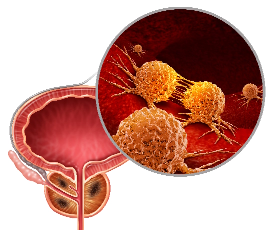
Cancer is a disease in which cells in an area begin changing and multiplying out of control. The multiplying cells may from a lump of tissue (tumor). With time, the cancer cells destroy healthy tissue and may spread to other parts of the body.
Bladder cancer is strongly linked to cigarette smoking. The longer a person smokes, the greater that person’s chances of developing bladder cancer.
Bladder cancer is often detected at the cystoscopy performed when a patient complains of passing blood in the urine. Other symptoms include urinary frequency.

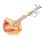
Cystoscopy
Cystoscopy
A thin telescope-like instrument called a cystoscope is inserted into the bladder through the urethra. This test allows the physician to visualise the inner lining of the bladder. If there are any abnormal looking areas, tiny samples of these areas are taken and sent for examination under the microscope by a pathologist (a biopsy). The results of a biopsy will usually be ready in a few days.
A flexible cystoscopy is usually performed in the clinic or day suite under local anesthesia. No fasting is required. A cystoscopy under anesthesia with a larger rigid instrument is usually performed if the surgeon has some other treatment in mind that he/she wants to perform at the same sitting. A rigid cystoscopy requires preparation for anesthesia and the patient to be fasted overnight
See more
To determine the best way to treat bladder cancer, your doctor checks
 The cancer STAGE
How deep the cancer has grown and where it has spread
The cancer GRADE
How aggressive the cancer looks and is likely to behave
The cancer STAGE is determined by performing an examination under anesthesia with a Transurethral Resection of Bladder Tumor (TURBT) and a CT scan of the pelvis. Studying the specimens obtained from the TURBT will tell us how deep the tumor has grown into the bladder wall. The CT will look for signs of cancer spread out of the bladder.
The cancer GRADE is determined from the TURBT specimens. They determine how aggressive the tumor can be and will determine the need for adjunctive treatment. This involves several visits to the doctor’s office where the bladder is applied with anti cancer medication.
Transurethral Resection of Bladder Tumor(TURBT)
Transurethral Resection of Bladder Tumor(TURBT)
Transurethral resection of bladder: This picture shows a “resectoscope” being inserted into the bladder through the penis. The tumor is then removed using a loop with cutting current.

CT Scan
CT Scan
An abdominal CT scan involves taking X-ray images of the abdomen from many angles. The X-ray beams are detected by the scanner and analyzed by a computer. The computer reconstructs the data into a picture of the body area being scanned. These images can be viewed on a monitor or reproduced as films.
The CT scanner is a free-standing machine with a large hole in the center. The patient lies on a narrow table that slides into the hole. Patients who have difficulty with enclosed spaces such as those found with some MRI scanners do not usually have a problem here.The actual scan time is usually about two minutes, although the entire procedure usually takes much longer.
A dye may be injected into a vein to better evaluate certain diseases and organs. When doing a quick screening for stones, dye injection is usually not necessary. A CT Scan offers several advantages over the IVP:
It is able to show the "meat" of the kidneys, where the kidney cancers and lumps are often situated. This is not seen in the IVP which only shows the inner outline of the urinary tract. This is why, if a lump is suspected on the IVP, fine CT sectioning of the kidney is performed to grade the likelihood of a lump being cancerous.
If a patient's symptoms are not very specific, CT allows a general "screening" of other abdominal organs in addition to the urinary tract.
See more
Bladder cancer begins in the lining of the bladder and often doesn’t grow beyond that layer. (Superficial Bladder Cancer). At this stage, it is eminently curable with a Transurethral Resection of Bladder Tumor (TURBT) with or without adjunctive treatment.
If the cancer has invaded the deeper layers of the bladder (Invasive Bladder Cancer), treatment will either involve removing the bladder (Radical Cystectomy) or radiotherapy to the bladder.
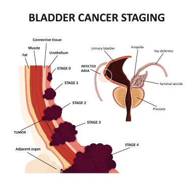
Superficial Bladder Cancer (TIS, TA or T1)
Cancer involving only the superficial lining of the bladder. This type of cancer is eminently curable with a TURBT. In some cases where the cancer is large, multiple or has minute involvement of the surrounding areas, further treatment is needed in the form of a course of chemotherapy instilled into the bladder.
Invasive Bladder Cancer (T2 and beyond)
If the bladder cancer has involved the muscle layers of the bladder or gone beyond to the surrounding fat or organs, it is called “invasive “ bladder cancer. This means that cure cannot be achieved just by TURBT alone. If the invasive cancer is still confined to the bladder only, treatment is by radical cystectomy or radiotherapy. If it has also involved other areas, you will usually be referred to an oncologist for chemotherapy.
Radical Cystectomy
Bladder tumors that have invaded the deeper layers but not gone beyond the bladder walls to its surroundings (T2) have a fighting chance of being cured if there are no other remote areas of involvement.
A radical cystectomy is a major operation to remove the bladder and surrounding tissue. The ureters will then have to be relocated into a loop of bowel that acts as a conduit, carrying it externally to a collection device. In suitable candidates, the bowel can be fashioned into a “bladder” like reservoir and stitched into the original bladder location. The patient then learns to void using the new “bladder”.