Hematuria

Hematuria is the abnormal presence of blood in the urine. There are two forms of hematuria. When the urine is visibly pink or red, it's called "gross hematuria." When no blood is visible in the urine, but detected by lab tests only, it is "microscopic hematuria".

Causes of Hematuria

Blood in the urine has many causes and can come from anywhere along the urinary tract.
The different causes of hematuria varies from:

Infections

Tumors

Kidney Disease

Trauma

These causes can lead to blood being seen or detected in the urine. Blood in the urine is therefore sometimes a warning signal and a thorough evaluation is necessary. If the cause is something serious, a physician wants to diagnose it early and begin proper treatment.

In some cases, no cause seems obvious even after a thorough evaluation. This occurs in as many as 50 percent of patients with microscopic hematuria and is known as ,”idiopathic hematuria”. These patients should be followed every six to 12 months with a repeat urine analysis and examination to see if things change.

Urinary Tract

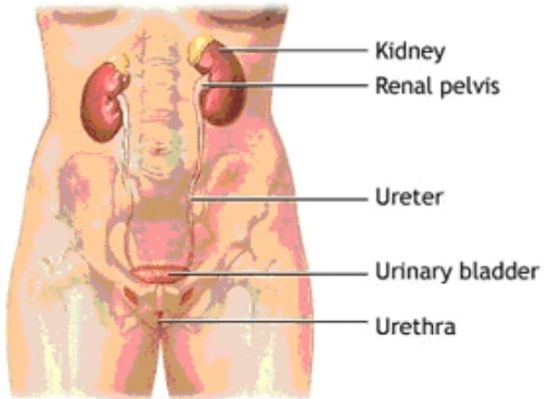
The urinary tract is made up of the kidneys, the ureters, the bladder, and the urethra.

Kidneys

The kidneys are a pair of bean-shaped organs located near the middle of the back. Each kidney filters waste products from the blood to form urine. Urine collects in tubules that eventually drain into the renal pelvis which continues as the ureter.

Ureters

The ureters are tubes that convey the urine from the kidney to the bladder. Muscles in the ureter walls constantly tighten and relax to force urine downward away from the kidneys. If this passage is blocked by a stone or by disease, kidney infection can develop. The area where the ureters enter the bladder is called the trigone. Valves in this region prevent the backing up of urine (reflux) into the kidneys.

Bladder

The urinary bladder is a hollow, muscular balloon that stores urine from the kidneys until it is an appropriate time to void. The bladder narrows down to an opening called the bladder neck. The bladder neck is surrounded by muscles called the internal sphincter which tighten around the urethra to prevent urine from leaking as the bladder fills. The bladder expels urine out of the body via the urethra.

Nerves in the bladder tell you when it is time to empty your bladder. When the volume of urine in the bladder reaches a certain capacity, the brain sends impulses to the internal sphincter to relax and to the bladder wall muscles to contract and expel urine. When all the signals occur in the correct order, normal urination occurs The bladder can usually hold up to 300-400ml of urine comfortably for 2-5 hours. Most people pass urine about 6-8 times a day.

Urethra and urethral sphincters

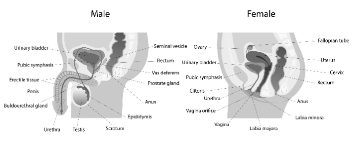
The urethra is the tube that passes urine from the bladder out of the body. The female urethra starts at the bladder neck and exits the body directly in front of the vaginal opening.

In men, the urethra is roughly 8 to 9 inches long and extends from the bladder neck to the end of the penis. The male urethra is composed of three parts: prostatic, membranous, and spongy. The prostatic urethra is the widest part of the tube and passes through the prostate gland.

Circular muscles called the external urethral sphincter also help keep urine from leaking. The sphincter muscles close tightly like a rubber band around the opening of the bladder into the urethra. This sphincter re-inforces the internal sphincter and is under voluntary control. This is one of the parts you learn to strengthen when you are taught pelvic floor exercises.

Male Reproductive System

Female lower urinary tract

The female bladder lies in front of the uterus (womb) and behind the pubic bone. The female bladder, bladder neck and urethra is supported by the pelvic muscles at the base like a hammock. Disruption, damage or loss of strength at this hammock can lead to pelvic organ prolapse and /or stress incontinence.

MEDICAL TERMS

PELVIC FLOOR MUSCLE TRAINING

Perhaps the best first-line approach for any form of incontinence is a combination of Kegel exercises and bladder training. In one study, women who used this combination approach experienced an average 50% reduction in incontinence episodes, with nearly 40% of them achieving complete continence. It was equally effective for urge, stress, or mixed incontinence.

Studies also reported that between 50% and 75% of patients who perform only pelvic floor exercises experience a substantial improvement in their symptoms, including elderly people who have had the problem for years.

Kegel’s or Pelvic floor muscle exercises are designed to strengthen the muscles of the pelvic floor that support the bladder and close the sphincters. Dr. Kegel first developed these exercises to assist women before and after childbirth, but they are very useful in helping to improve continence for both men and women. Studies indicate that such exercises are very effective, even for men recovering from surgery for prostate cancer.

Kegel exercises are particularly useful for the following:

Stress incontinence. Some experts believe that Kegel exercises should be the primary treatment for stress incontinence.

Urge incontinence. They can also be helpful for urge incontinence in cases that are not caused by nerve damage. In one study, 85% of women reported satisfaction with this program.

At Pearllyn Quek Urology, your pelvic floor contraction efforts will be assessed and graded before starting your PFMT program. This grading, along with your clinical progress will be reviewed over 3 months.

Rating

Contraction

Duration

0

None

None

1

Flicker

None

2

Slight Squeeze

Not Sustained

3

Moderate Squeeze

2 to 3 Secs.

4

Good Squeeze

3 to 5 Secs.

5

Strong Squeeze

More than 5 Secs.

Grading of the pelvic floor muscle using the modified Oxford Scale(Laycock 1994)

It will usually take 3 months before a patient sees significant improvement.

Incontinence will return to its original severity if these exercises are discontinued, so commitment to the program must be high and possibly life-long.

These bar charts show how the majority of our patients improve from Grades 0-3 (poor or unsustained contractions) to grades 3-5 (dark blue bars) at the end of our 3 months program.

Dark blue bars – After PFMT
Light blue bars – Before PFMT
Grade 1 = Mild, occasional stress incontinence that does not require surgery
Grade 2 = Moderate stress incontinence often requiring surgery
Grade 3 = Severe daily stress incontinence definitely requiring surgery

This bar chart shows how the majority of patients undergoing our pelvic floor training are cured (dry) or improved (shifted to Gd 1) compared to before (light blue bars)

Return

We carry out a standardized evaluation of patients with hematuria consisting of the following:

1

Urine Cytology

A sample of urine is analysed under the microscope by a pathologist for cancer cells. If positive, it indicates a cancer is growing within the urinary tract. No conclusion can be drawn if negative as the sensitivity of this test ranges from 20% for the less aggressive looking cancers to 80% for the more aggressive ones.

See more
2

Urine Culture and Sensitivity

A sample of urine is tested for the presence of clinically significant amounts of bacteria. If positive, it indicates a urinary tract infection (UTI). A panel of antibiotics is then tested against the bacteria cultured to determine which works and which doesn't. This test can be negative in up to 30% of females with UTI if they are also experiencing urinary frequency at the time of infection.

See more
3

Blood tests and urinary protein levels

4

IVP (Intravenous Pyelogram) or equivalent

This is an xray examination that studies the outlines of the urinary tract (kidney, ureter and bladder). After xray dye is injected into a vein, serial xrays of the abdominal area follows the dye as it passes through the urinary tract. IVPs can demonstrate urinary stones, blockage and abnormalities in the inner lining of the urinary tract. It also gives some indication of any difference in kidney function between the right and left kidneys. It does not show up the "meat" or parenchymal area of the kidney well, therefore an ultrasound or CT scan is required to further elucidate any kidney masses suspected on an IVU.

See more
5

Cystoscopy

A thin telescope-like instrument called a cystoscope is inserted into the bladder through the urethra. This test allows the physician to visualise the inner lining of the bladder. If there are any abnormal looking areas, tiny samples of these areas are taken and sent for examination under the microscope by a pathologist (a biopsy). The results of a biopsy will usually be ready in a few days.


A flexible cystoscopy is usually performed in the clinic or day suite under local anesthesia. No fasting is required. A cystoscopy under anesthesia with a larger rigid instrument is usually performed if the surgeon has some other treatment in mind that he/she wants to perform at the same sitting. A rigid cystoscopy requires preparation for anesthesia and the patient to be fasted overnight.

See more

Treatment

Most hematuria can be evaluated on a non-urgent basis. Treatment depends on the cause